Brainstem Connectomes
Read More

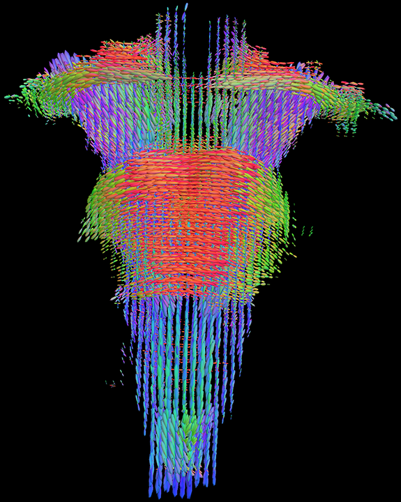
FOD-based, topography-preserving tractography techniques from NICR for the modeling of human connectomes
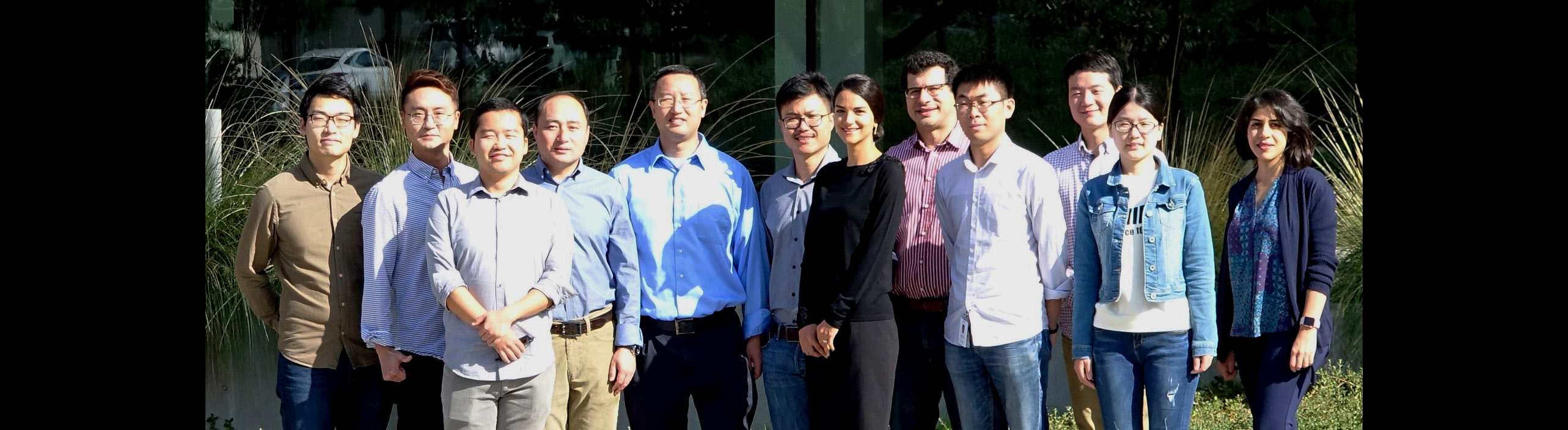
The Neuro Image Computing Research (NICR) group is led by Dr. Yonggang Shi at the Mark and Mary Stevens Neuroimaging and Informatics Institute of USC. The main goal of the NICR group is to provide innovative solutions for challenging computational problems in brain imaging and related fields. In our research, we focus on developing novel 3D shape analysis and connectome modeling techniques to achieve more accurate and reliable measurements of brain anatomy and function. We are also interested in generalizing these computational tools to other medical imaging domains such as retinal imaging. Through collaborations with leading experts in clinical and neuroscience research, we are committed to advancing brain disorder research with our image computing techniques. To learn more about our research, please take a look at members of our group, their exciting projects, and papers.
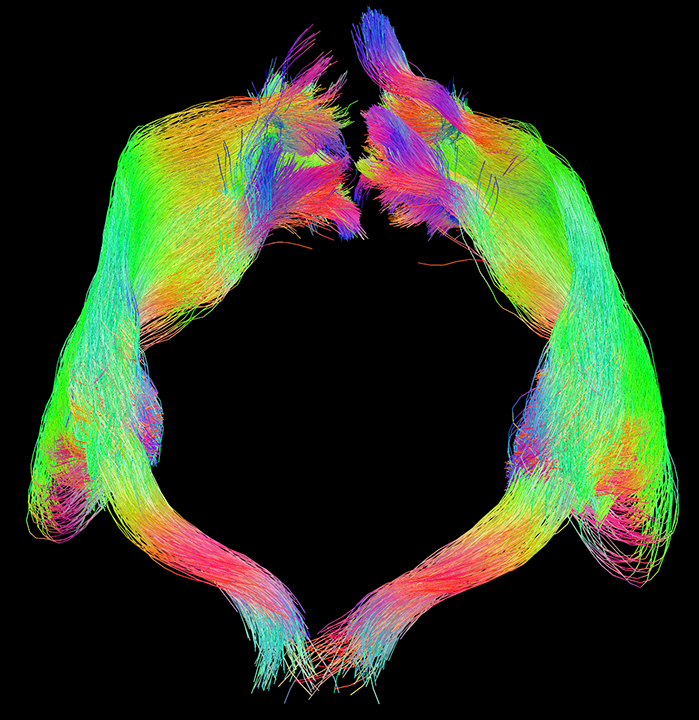
Increasing evidence from various neuropathology studies and recently updated Braak staging suggested that the earliest tau pathology of Alzheimer’s disease (AD) may occur in the brainstem nuclei and propagate to trans-entorhinal cortical regions. In this project, we will develop the enabling techniques for the mapping of brainstem pathways using multi-shell diffusion MRI, which can provide connectivity measures to complement cortical tau burdens from PET imaging. With the novel brainstem atlases and associated software tools developed in this project, we will be able to examine the relation of brainstem and cortical atrophy during the disease course of AD. In addition, the brainstem connectomes will enable the in vivo mapping of the connectivity changes in neurotransmitter-specific pathways and their relation to behavior symptoms in AD patients.
Grant Support: NIA R01AG056573.
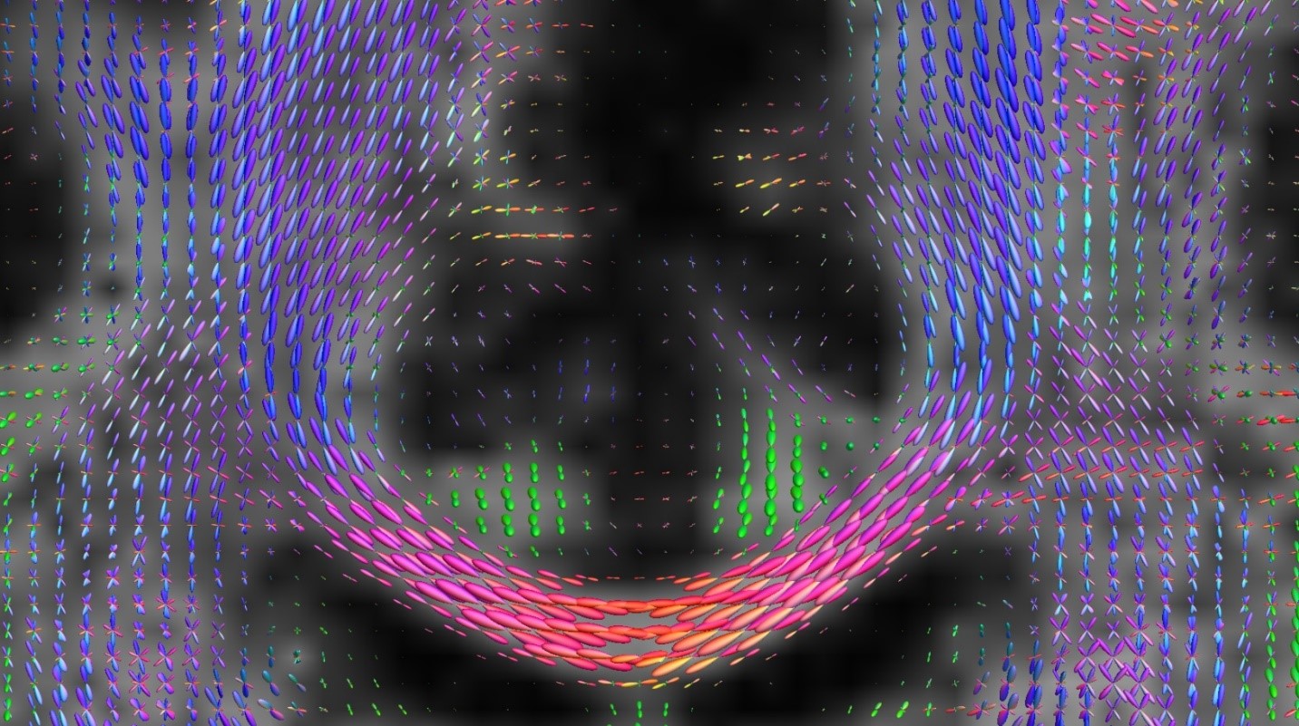
For the in vivo investigation of brain connectome, diffusion MRI (dMRI) is an important tool as it provides highly sensitive imaging markers and allows the examination of connection paths via tractography. With the success of the Human Connectome Project (HCP), high-resolution, multi-shell diffusion imaging is emerging as the standard approach for dMRI data acquisition in connectome studies. In this project we will develop a suite of novel computational tools that jointly estimate fiber orientation distributions (FOD) and compartmental parameters. We will also develop novel, topography-preserving tractography and filtering techniques, and their validation with mouse tracer injection data.
Grant Support: NIBIB R01EB022744, NIA U01AG051218, NIA P50AG05142.
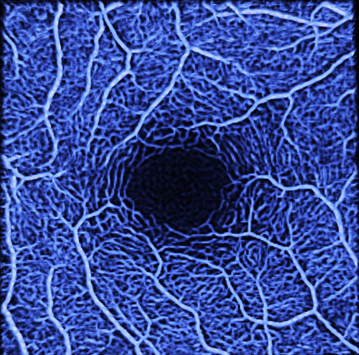
How does the precise topography of eye disease map onto changes in brain? Is acquired retinal damage related to local effects upon the structure and function of the CVP? Are any of these effects of vision loss on the CVP reversible? We propose to answer these questions, which we believe are crucial to the on-going efforts in developing sightrestoration treatments, by combining advanced retinal imaging with the brain-mapping techniques developed in the Human Connectome Project (HCP), using novel yet robust analytical methods.
Grant Support: NEI U01EY025864
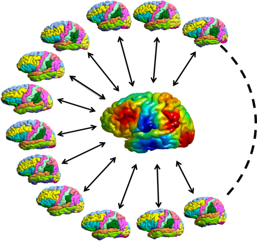
We focus on developing novel methods for surface reconstruction and mapping that includes both cortical and sub-cortical structures. Using fast-level-set methods and Laplace-Beltrami spectral analysis, we developed a novel framework of surface reconstruction for both cortical and sub-cortical structures. For the mapping brain structures, we developed a general approach based on Riemannian Metric Optimization on Surfaces (RMOS) that can compute general diffeomorphisms and match connectivity patterns across different brains.
Grant Support: NIBIB P41EB015922, NIBIB K01EB013633, NIA P01AG026572, Foundation of the ASNR.
In this project, we collaborate with Dr. Amir Kashani of the Roski Eye Institute of USC to develop 3D metrics for the automated analysis of OCT and OCTA data and apply them for the early diagnosis of diabetic retinopathy and neurovascular diseases. We will translate and adapt image computing tools from our brain imaging research to 3D retinal vasculature modeling and analysis.
Grant Support: NEI R21EY027879, NINDS UH2NS100614.
Here we list 10 papers most related to our projects. For a more complete list of our publication, please find it on the Google Scholar link of individual members.
Nie X, Ruan J, Otaduy MCG, Grinberg LT, Ringman J, Shi Y. Surface-Based Probabilistic Fiber Tracking in Superficial White Matter. IEEE Trans Med Imaging. 2024 Mar;43(3):1113-1124. PMID: 37917515; PMCID: PMC10917128.
Xia Y, Shi Y. Diffusion MRI Harmonization via Personalized Template Mapping. Hum Brain Mapp. 2024 Apr; 45(5):e26661. PMID: 38520363; PMCID: PMC10960558.
Qiao Y, Shi Y. Unsupervised Deep Learning for FOD-Based Susceptibility Distortion Correction in Diffusion MRI. IEEE Trans Med Imaging. 2022 May;41(5):1165-1175. PMID: 34882551; PMCID: PMC9177803.
Aydogan DB, Shi Y. Parallel Transport Tractography. IEEE Trans Med Imaging. 2021 Feb;40(2):635-647. PMID: 33104507; PMCID: PMC7931442.
Zhang J, Qiao Y, Sarabi MS, Khansari MM, Gahm JK, Kashani AH, Shi Y. 3D Shape Modeling and Analysis of Retinal Microvasculature in OCT-Angiography Images. IEEE Trans Med Imaging. 2020 May;39(5):1335-1346. PMID: 31647423; PMCID: PMC7174137.
Aydogan DB, Jacobs R, Dulawa S, Thompson SL, Francois MC, Toga AW, Dong H, Knowles JA, Shi Y. When tractography meets tracer injections: a systematic study of trends and variation sources of diffusion-based connectivity. Brain Struct Funct. 2018 Jul;223(6):2841-2858. PMID: 29663135; PMCID: PMC5997540.
Gahm JK, Shi Y; Alzheimer's Disease Neuroimaging Initiative. Riemannian metric optimization on surfaces (RMOS) for intrinsic brain mapping in the Laplace-Beltrami embedding space. Medical Image Analysis. 2018 May;46:189-201. PMID: 29574399; PMCID: PMC5910235.
Tang Y, Sun W, Toga AW, Ringman JM, Shi Y. A probabilistic atlas of human brainstem pathways based on connectome imaging data. Neuroimage. 2018 Apr 1;169:227-239. PMID: 29253653; PMCID: PMC5856609.
Tran G, Shi Y. Fiber Orientation and Compartment Parameter Estimation From Multi-Shell Diffusion Imaging. IEEE Trans Med Imaging. 2015 Nov;34(11):2320-32. PMID: 25966471; PMCID: PMC4657863.
Shi Y, Lai R, Toga AW; Alzheimer's Disease Neuroimaging Initiative. Cortical surface reconstruction via unified Reeb analysis of geometric and topological outliers in magnetic resonance images. IEEE Trans Med Imaging. 2013 Mar;32(3):511-30. PMID: 23086519; PMCID: PMC3785796.